


In collaboration with outside experts, we have explored the ways in which brain imaging may illuminate and extend the findings from our regular testing. Funding for much of this research has been provided by the Johnson O’Connor Research Support Corporation. Here is a brief summary of where it all is leading. This research won’t provide direct guidance yet, but it does provide substantiating evidence for the traits we have identified and measured over the years.
The Foundation’s founder and namesake, Johnson O’Connor, had an abiding interest in the biological substrate of individual differences in aptitudes. In late 2006, at a professional research conference, our President, David Ransom, discussed with Dr. Richard Haier, a leading researcher on brain imaging and intelligence, the possibility of relating the volumes of defined brain areas measured with structural magnetic resonance imaging (sMRI) to performance on Johnson O’Connor aptitude tests.
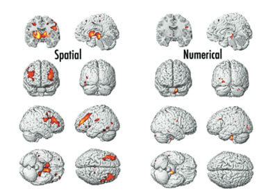
In the spring of 2007 Dr. Haier agreed to work on such a study, and in conjunction with Mt. Sinai Medical Center in New York, to conduct sMRI scans of 40 Foundation examinees under the supervision of Dr. Cheuk Tang. Dr. Haier used voxel-based morphometry to identify various brain areas and measure the volume of gray and white matter in each area. The accompanying figures below show brain areas that are related to indexes derived from spatial and numerical aptitudes. As can be seen, there is a stark contrast in the two types of thinking in relation to brain areas highlighted. This was just the beginning of intriguing basic research that we hope will lead to breakthroughs in what we know about the brain and ultimately, how individual examinees can benefit from this new approach.
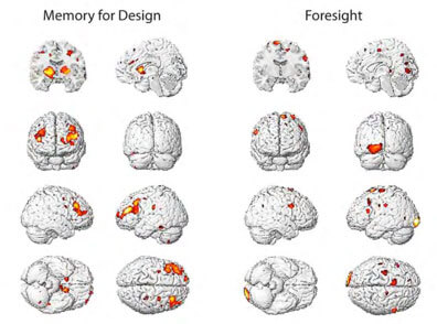
Further research demonstrated a strong contrast using scores directly derived from two very different aptitudes. The areas displayed at right in red and yellow colors indicate significant positive correlations between test performance and densities in the given brain areas. Foresight is related to a number of brain areas, including a sizable area in the left occipital lobe. Memory for Design is distinctly different, with several relatively large areas, including in the left and right frontal lobes. Memory for Design, befitting its correlations to spatial thinking, shows similarity to the Spatial brain images above. See the papers by Haier et al. listed on the Publications and Presentations page.
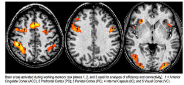
Dr. Tang and Dr. Haier tested the efficiency hypothesis—that a brain shows less activation when working more successfully—on the fMRI data collected while examinees performed a working-memory task. They found that higher structural visualization was indeed related to more efficient processing (lesser activation) in the right prefrontal cortex and right parietal cortex (note that spatial processing tends to be localized in the right hemisphere in most persons).
In late 2010 we made arrangements to collaborate with the MIND Institute for brain imaging, which is affiliated with the University of New Mexico, to conduct a study in which magnetoencephalography (MEG) imaging is used to create a continuous picture (that is, a video) of brain functioning in examinees while they take alternate items for several Foundation tests. Our alternate versions needed to be formatted to fit the constraints examinees are under during MEG imaging. The head and body must remain very still, with only fingers free to manipulate a computer track ball while watching a cursor move on a screen with test questions projected. The principal investigator for this study is Haier’s long-time friend and collaborator Dr. Rex Jung, of the University of New Mexico.

We have continued working with Dr. Jung and his associates on other studies, including a study of twins. (Also see our prior twin study presented as a separate research topic). In 2014 their paper Subcortical Correlates of Individual Differences in Aptitude was published in the online journal PLoS ONE. Here is one figure from this article.

Our previous scholarly work has continued to receive attention. Our 2010 article with Dr. Richard Haier and his associates in BMC Research Notes has now been viewed as of June 2015 over 10,000 times, while our 2012 article by Dr. David Schroeder and others in BMC has been viewed some 900+ times. According to Google Scholar, the 2010 Haier et al. article has been cited in 10 scholarly journal articles and books, and our 2009 article with Haier and others in Intelligence has been cited 44 times. In addition, our 2010 article with Dr. Cheuk Tang and others in Intelligence has been cited in 34 articles and books.